| Citation: |
DENG Jianhao, LIU Li, SHU Chan, et al. Transcriptome analysis of Drosophila S2 cells after infection of Listeria monocytogenes, L. grayi or L. welshimeri[J]. Journal of South China Agricultural University, 2025, 46(1): 12-24. DOI: 10.7671/j.issn.1001-411X.202312026

|
To compare the gene expression patterns in S2 cells after infection of Listeria monocytogenes, L. grayi, and L. welshimeri using Drosophila as a host, and provide a research basis for exploring the innate immune defense mechanisms of the host and the differential pathogenic mechanisms of different Listeria strains.
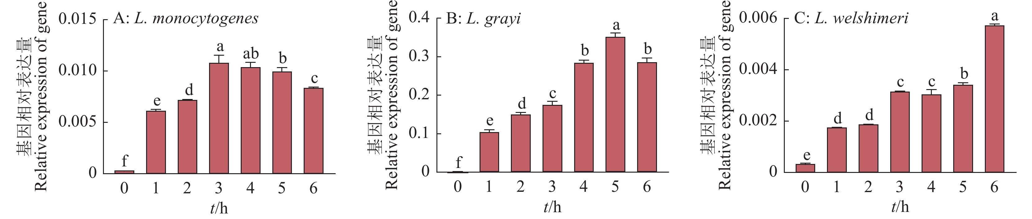
After infection of L. monocytogenes, L. grayi or L. welshimeri for 3 h, the mRNA expression profiles in Drosophila S2 cells were detected using transcriptome sequencing technology. Analysis of differentially expressed genes (DEGs) was performed using bioinformatics tools of GO annotation and KEGG analysis. Finally, the mRNA levels of five antimicrobial peptide genes associated with the Toll and Imd signaling pathways were validated using qRT-PCR technology.
After infection of L. monocytogenes, L. gray or L. welshimeri, there were 18 DEGs in common, as well as 104, 28, and 33 unique DEGs in the Drosophila S2 cells respectively. The DEGs commonly existed in three Listeria infected groups were annotated in GO terms including metabolic process, cellular process, organelle, response to stimulus, immune system process and etc. The unique DEGs existed in L. monocytogenes infected group were annotated in GO terms such as biological adhesion, presynaptic process involved in chemical synaptic transmission, negative regulation of biological process, nucleic acid binding transcription factor activity, signal transducer activity, and etc. The unique DEGs in L. grayi infected group were annotated in GO terms such as multi-organism process and extracellular regions, while the unique DEGs in L. welshimeri infected group were annotated in GO terms such as developmental processes and membrane-enclosed lumen. The KEGG analysis results showed that the DEGs of three Listeria infected groups were enriched in the Toll/Imd signaling pathways. The quantitative results of qRT-PCR validation of five antimicrobial peptide genes related to Toll and Imd signaling pathways were consistent with the transcriptomic results.
The expression patterns in S2 cells affected by the infection of these three Listeria species exhibited some similarities but also differences. The variation of the signaling pathways may be related to the virulence of these Listeria species in S2 cells.
| [1] |
COLLINS M D, WALLBANKS S, LANE D J, et al. Phylogenetic analysis of the genus Listeria based on reverse transcriptase sequencing of 16S rRNA[J]. International Journal of Systematic Bacteriology, 1991, 41(2): 240-246. doi: 10.1099/00207713-41-2-240
|
| [2] |
何冬梅, 邓峰, 赖蔚苳, 等. 单核细胞增生李斯特菌生物学研究进展[J]. 华南预防医学, 2006, 32(6): 26-29.
|
| [3] |
柴文琴. 李斯特菌属不同种型在模拟食品加工环境相关胁迫下的存活率与耐药性研究[D]. 杭州: 浙江大学, 2020.
|
| [4] |
KAPTCHOUANG TCHATCHOUANG C D, FRI J, DE SANTI M, et al. Listeriosis outbreak in South Africa: A comparative analysis with previously reported cases worldwide[J]. Microorganisms, 2020, 8(1): 135. doi: 10.3390/microorganisms8010135
|
| [5] |
CHARLIER C, PERRODEAU É, LECLERCQ A, et al. Clinical features and prognostic factors of listeriosis: The MONALISA national prospective cohort study[J]. The Lancet Infectious Diseases, 2017, 17(5): 510-519. doi: 10.1016/S1473-3099(16)30521-7
|
| [6] |
中华人民共和国国家卫生健康委员会, 国家市场监督管理总局. 食品安全国家标准 预包装食品中致病菌限量: GB 29921—2021[S]. 北京: 中国标准出版社, 2021.
|
| [7] |
中华人民共和国家卫生和计划生育委员会. 食品安全国家标准 食品中致病菌限量: GB 29921—2013[S]. 北京: 中国标准出版社, 2014.
|
| [8] |
VÁZQUEZ-BOLAND J A, KUHN M, BERCHE P, et al. Listeria pathogenesis and molecular virulence determinants[J]. Journal of Personalized Medicine, 2001, 14(3): 584-640.
|
| [9] |
李钊, 刘阳泰, 李卓思, 等. 单增李斯特菌毒力因子及调控机制研究进展[J]. 食品与发酵工业, 2024, 50(11): 327-335.
|
| [10] |
IRETON K, MORTUZA R, GYANWALI G C, et al. Role of internalin proteins in the pathogenesis of Listeria monocytogenes[J]. Molecular Microbiology, 2021, 116(6): 1407-1419. doi: 10.1111/mmi.14836
|
| [11] |
ZHOU H, COVENEY A P, WU M, et al. Activation of both TLR and NOD signaling confers host innate immunity-mediated protection against microbial infection[J]. Frontiers in Immunology, 2019, 9: 3082. doi: 10.3389/fimmu.2018.03082
|
| [12] |
MITCHELL G, CHENG M I, CHEN C, et al. Listeria monocytogenes triggers noncanonical autophagy upon phagocytosis, but avoids subsequent growth-restricting xenophagy[J]. Proceedings of the National Academy of Sciences of the United States of America, 2017, 115(2): E210-E217.
|
| [13] |
CHEN G Y, PENSINGER D A, SAUER J D. Listeria monocytogenes cytosolic metabolism promotes replication, survival, and evasion of innate immunity[J]. Cellular Microbiology, 2017, 19(10): e12762-e12771. doi: 10.1111/cmi.12762
|
| [14] |
GABALLA A, GUARIGLIA-OROPEZA V, WIEDMANN M, et al. Cross talk between SigB and PrfA in Listeria monocytogenes facilitates transitions between extra- and intracellular environments[J]. Microbiology and Molecular Biology Reviews, 2019, 83(4): e00034-19.
|
| [15] |
BIERNE H, HAMON M, COSSART P. Epigenetics and bacterial infections[J]. Cold Spring Harbor Perspectives in Medicine, 2012, 2(12): a010272
|
| [16] |
MINÁROVITS J. Microbe-induced epigenetic alterations in host cells: The coming era of patho-epigenetics of microbial infections: A review[J]. Acta Microbiologica et Immunologica Hungarica, 2009, 56(1): 1-19. doi: 10.1556/AMicr.56.2009.1.1
|
| [17] |
HAMON M A, COSSART P. Histone modifications and chromatin remodeling during bacterial infections[J]. Cell Host & Microbe, 2008, 4(2): 100-109.
|
| [18] |
COSTA A C, PINHEIRO J, REIS S A, et al. Listeria monocytogenes interferes with host cell mitosis through its virulence factors InlC and ActA[J]. Toxins, 2020, 12(6): 411-424. doi: 10.3390/toxins12060411
|
| [19] |
袁江北. 定量蛋白质组学揭示免疫细胞对李斯特菌感染的防御机制[D]. 重庆: 重庆大学, 2020.
|
| [20] |
IMLER J L. Overview of Drosophila immunity: A historical perspective[J]. Developmental & Comparative Immunology, 2014, 42(1): 3-15.
|
| [21] |
GOTTAR M, GOBERT V, MICHEL T, et al. The Drosophila immune response against Gram-negative bacteria is mediated by a peptidoglycan recognition protein[J]. Nature, 2002, 416: 640-644. doi: 10.1038/nature734
|
| [22] |
SCHMID M W, NG E Y W, LAMPIDIS R, et al. Evolutionary history of the genus Listeria and its virulence genes[J]. Systematic and Applied Microbiology, 2005, 28(1): 1-18. doi: 10.1016/j.syapm.2004.09.005
|
| [23] |
PIZARRO-CERDA J, COSSART P. Listeria monocytogenes: Cell biology of invasion and intracellular growth[J]. Microbiology Spectrum, 2018, 6(6). doi: 10.1128/microbiolspec.GPP3-0013-2018.
|
| [24] |
FREITAG N E, PORT G C, MINER M D. Listeria monocytogenes: From saprophyte to intracellular pathogen[J]. Nature Reviews Microbiology, 2009, 7: 623-628. doi: 10.1038/nrmicro2171
|
| [25] |
刘甜, 罗开珺. 果蝇Toll和IMD信号通路中的功能结构域[J]. 环境昆虫学报, 2011, 33(3): 388-395. doi: 10.3969/j.issn.1674-0858.2011.03.015
|
| [26] |
LEULIER F, RODRIGUEZ A, KHUSH R S, et al. The Drosophila caspase Dredd is required to resist gram-negative bacterial infection[J]. EMBO Reports, 2000, 1(4): 353-358. doi: 10.1093/embo-reports/kvd073
|
| [27] |
BALDWIN D N, VANCHINATHAN V, BROWN P O, et al. A gene-expression program reflecting the innate immune response of cultured intestinal epithelial cells to infection by Listeria monocytogenes[J]. Genome Biology, 2002, 4(1): R2. doi: 10.1186/gb-2002-4-1-r2
|
| [28] |
吴坤钟, 李荣松, 曹阳, 等. 英诺克李斯特菌侵染果蝇S2细胞后的转录组分析[J]. 华南农业大学学报, 2020, 41(5): 17-26. doi: 10.7671/j.issn.1001-411X.202001023
|
| [29] |
王旭. 基于比较基因组学和转录组学技术揭示单增李斯特菌毒力因子的研究[D]. 上海: 上海海洋大学, 2016.
|
| [30] |
PIZARRO-CERDÁ J, COSSART P. Subversion of cellular functions by Listeria monocytogenes[J]. Journal of Pathology, 2006, 208(2): 215-223. doi: 10.1002/path.1888
|
| [31] |
KRYPOTOU E, SCORTTI M, GRUNDSTRÖM C, et al. Control of bacterial virulence through the peptide signature of the habitat[J]. Cell Reports, 2019, 26(7): 1815-1827. e5.
|
| [32] |
SAISAWANG C, WONGSANTICHON J, KETTERMAN A J. A preliminary characterization of the cytosolic glutathione transferase proteome from Drosophila melanogaster[J]. Biochemical Journal, 2012, 442(1): 181-190. doi: 10.1042/BJ20111747
|
| [33] |
XIE A, ODATE S, CHANDRAMOULY G, et al. H2AX post-translational modifications in the ionizing radiation response and homologous recombination[J]. Cell Cycle (Georgetown, Tex), 2010, 9(17): 3602-3610. doi: 10.4161/cc.9.17.12884
|
| [34] |
WU M H, ZHANG X D, WEI W, et al. CRISPR/Cas9 mediated genetic resource for unknown kinase and phosphatase genes in Drosophila[J]. Scientific Reports, 2020, 10: 7383. doi: 10.1038/s41598-020-64253-4
|
| [35] |
SCHERFER C, KARLSSON C, LOSEVA O, et al. Isolation and characterization of hemolymph clotting factors in Drosophila melanogaster by a pullout method[J]. Current Biology, 2004, 14(7): 625-629. doi: 10.1016/j.cub.2004.03.030
|
| [36] |
单颖. 单核细胞增生李斯特菌: 无菌斑马鱼感染模型及Mmp-9在抗细菌感染中的作用机制[D]. 杭州: 浙江大学, 2016.
|
Supported by: Beijing Renhe Information Technology Co., Ltd. 
 DownLoad: