
Distribution of IgA secreting cells in respiratory mucosa of chicken at different day-age
-
摘要:目的
了解鸡呼吸道黏膜不同时期的免疫状态。
方法选择海兰白鸡鸡胚(18、20 日龄)及不同日龄(1、4、7、14、21、35和56 日龄)雏鸡的鼻、喉、气管和肺脏样品,利用免疫组织化学方法研究IgA+分泌细胞的出现、定位分布及数量变化过程。
结果呼吸道各个器官黏膜中胚胎时期均没有IgA+细胞出现,鼻和肺脏中1日龄时出现,喉黏膜中4日龄时出现,而气管黏膜中7日龄时才出现。随着日龄增长,各器官呼吸道黏膜中IgA+细胞数量均逐渐增加。鼻黏膜以及肺脏初级支气管和次级支气管交叉处4日龄时较早地形成淋巴聚集物,喉黏膜中7日龄时形成,气管黏膜中35日龄时形成。在这些淋巴聚集物中IgA+细胞均主要分布于淋巴聚集物的外周。35日龄时,在鼻、喉和气管黏膜固有层以及上皮内均存在较多的IgA+细胞,从而能够更有效和直接地分泌SIgA,执行黏膜免疫功能。56日龄时,4种器官的呼吸道黏膜中IgA+细胞的数量达到峰值,并在黏膜底部形成生发中心,具有了黏膜相关性淋巴组织的典型特征,从而更有效地执行黏膜免疫功能。
结论鸡呼吸道黏膜中IgA+细胞的分布和数量均呈现日龄相关性变化,并且在35日龄时,鼻、喉、气管和肺脏中IgA+细胞数量和分布部位均达到一定规模,能够为呼吸道黏膜提供免疫保护。
Abstract:ObjectiveTo understand the immune status of chicken respiratory mucosa at different stages.
MethodThe embryo of Hy-line white chickens (18- and 20-day-old), and the nose, larynx, trachea and lung of chickens at different day-age (1-, 4-, 7-, 14-, 21-, 35- and 56-day-old) were selected in this study. The occurrence, location, distribution and quantity change of IgA+ secreting cells were studied by immunohistochemical method.
ResultThere was no IgA+ cell in the mucosa of each respiratory organ during the embryonic period. IgA+ cells were present in the nose and lung at 1-day-old age, laryngeal mucosa at 4-day-old age, and tracheal mucosa at 7-day-old age. The number of IgA+ cells in the respiratory mucosa of each organ increased gradually with the increase of age. The lymphoid aggregates were formed earlier in the nasal mucosa and the intersections of the primary and secondary bronchus in the lung at 4-day-old age, and formed in the laryngeal mucosa at 7-day-old age, in the tracheal mucosa at 35-day-old age. The IgA+ cells in these lymphoid aggregates were all mainly distributed on the periphery of the lymphoid aggregates. At 35-day-old age, there were more IgA+ cells in the mucosal lamina propria and the epithelium of the nose, larynx and trachea, and thus more effective and direct secretion of SIgA could be performed for mucosal immunity. At 56-day-old age, the number of IgA+ cells in the respiratory mucosa of the four organs reached a peak and germinal centers were formed at the mucosal bottom, which was the characteristic of mucosal associated lymphoid tissue, thus more effectively performed mucosal immune function.
ConclusionThe distribution and number of IgA+ cells in the respiratory mucosa of chickens show age-related changes, and the number and distribution of IgA+ cells in the nose, larynx, trachea and lung all reach a certain scale at 35-day-old, which can provide immune protection for the respiratory mucosa.
-
Keywords:
- chicken /
- respiratory mucosa /
- IgA+ secreting cells /
- distribution characteristic /
- mucosal immune
-
-
![]()
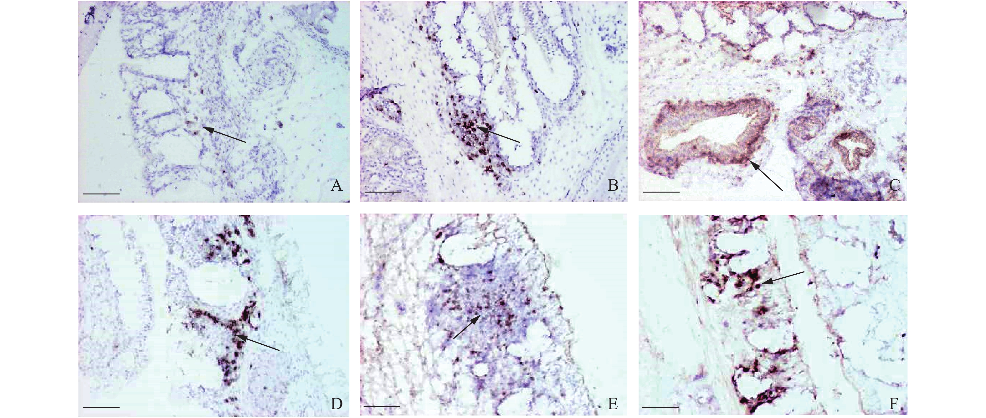
图 1 不同日龄鸡鼻黏膜中IgA+分泌细胞的分布情况
A:1日龄(箭头指向鼻后孔鼻腔侧黏膜下的IgA+细胞),B:4日龄(箭头指向鼻黏膜相关性淋巴组织中的IgA+细胞),C:4日龄(箭头指向鼻腺周围的IgA+细胞),D:7日龄(箭头指向鼻黏膜相关性淋巴组织中的IgA+细胞),E:21日龄(箭头指向鼻黏膜相关性淋巴组织中的IgA+细胞),F:35日龄(箭头指向柱状皱襞固有层中的IgA+细胞);标尺=100 μm
Figure 1. Distribution of IgA+ secreting cells in the nasal mucosa of chicken at different day-age
A: 1-day-old (arrow points to IgA+ cells in the submucosa of the nasal cavity in the posterior nasal foramen), B: 4-day-old (arrow points to IgA+ cells in the associated lymphoid tissue of nasal mucosa), C: 4-day-old (arrow points to IgA+ cells around the nasal glands), D: 7-day-old (arrow points to IgA+ cells in the associated lymphoid tissue of nasal mucosa), E: 21-day-old (arrow points to IgA+ cells in the associated lymphoid tissue of nasal mucosa), F: 35-day-old (arrow points to IgA+ cells in the columnar plica lamina propria); Bar = 100 μm
![]()
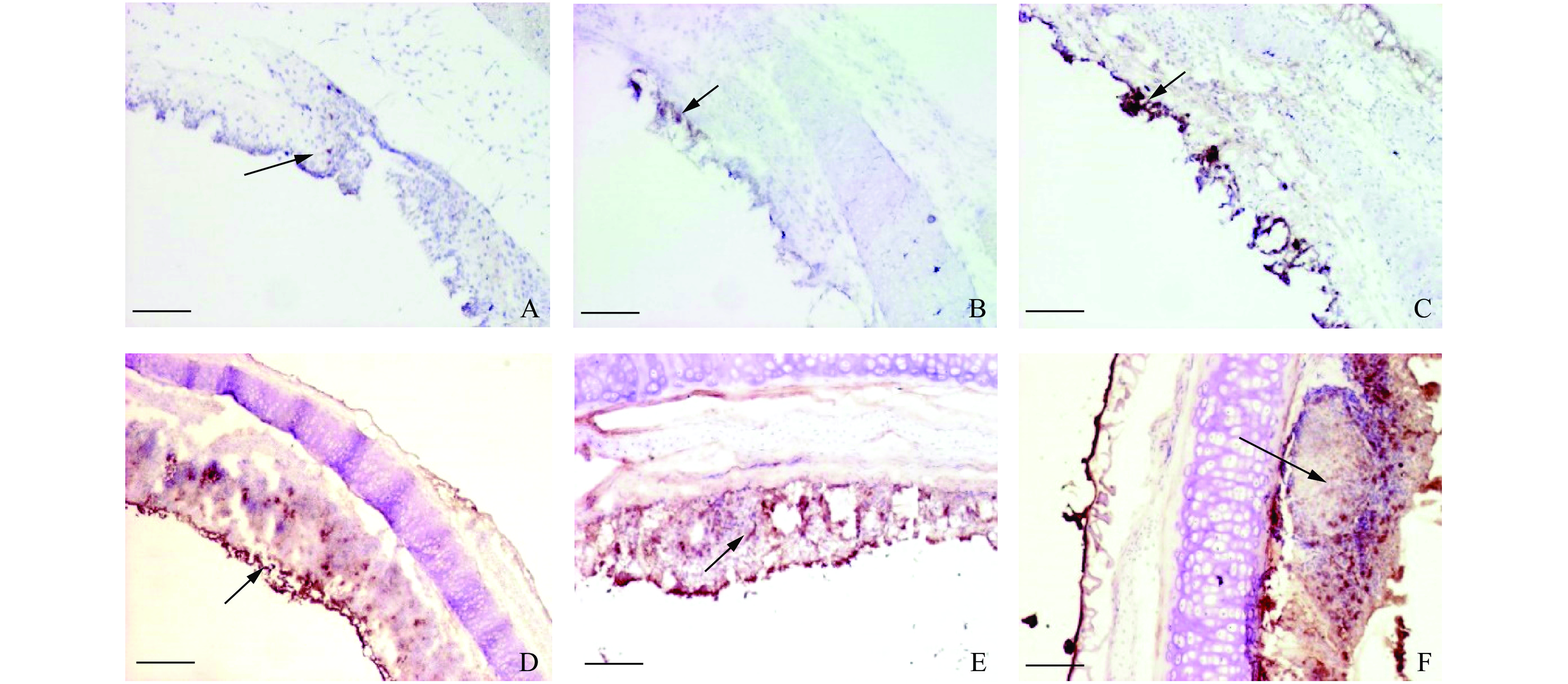
图 2 不同日龄鸡喉黏膜中IgA+分泌细胞的分布情况
A:4日龄(箭头指向喉正中黏膜嵴中的IgA+细胞),B:7日龄(箭头指向淋巴聚集物中的IgA+细胞),C:14日龄(细箭头指向喉口处喉腔侧的呼吸性假复层柱状纤毛上皮下形成的淋巴聚集物,粗箭头指向复层扁平上皮),D:21日龄(箭头指向黏膜上皮中的IgA+细胞),E:35日龄(箭头指向集中在淋巴聚集物外周的IgA+细胞),F:56日龄(箭头指向生发中心);标尺=100 μm
Figure 2. Distribution of IgA+ secreting cells in the laryngeal mucosa of chicken at different day-age
A: 4-day-old (arrow points to IgA+ cells in the mid-laryngeal mucosa cristae), B: 7-day-old (arrow points to IgA+ cells in the lymphoid aggregates), C: 14-day-old (thin arrow points to the lymphatic aggregates formed under respiratory pseudostratified columnar ciliated epithelium at the laryngeal opening, thick arrow points to the stratified flattened epithelium), D: 21-day-old (arrow points to IgA+ cells in the mucosal epithelium), E: 35-day-old (arrow points to IgA+ cells concentrated around the lymphoid aggregates), F: 35-day-old (arrow points to the germinal center); Bar = 100 μm
![]()
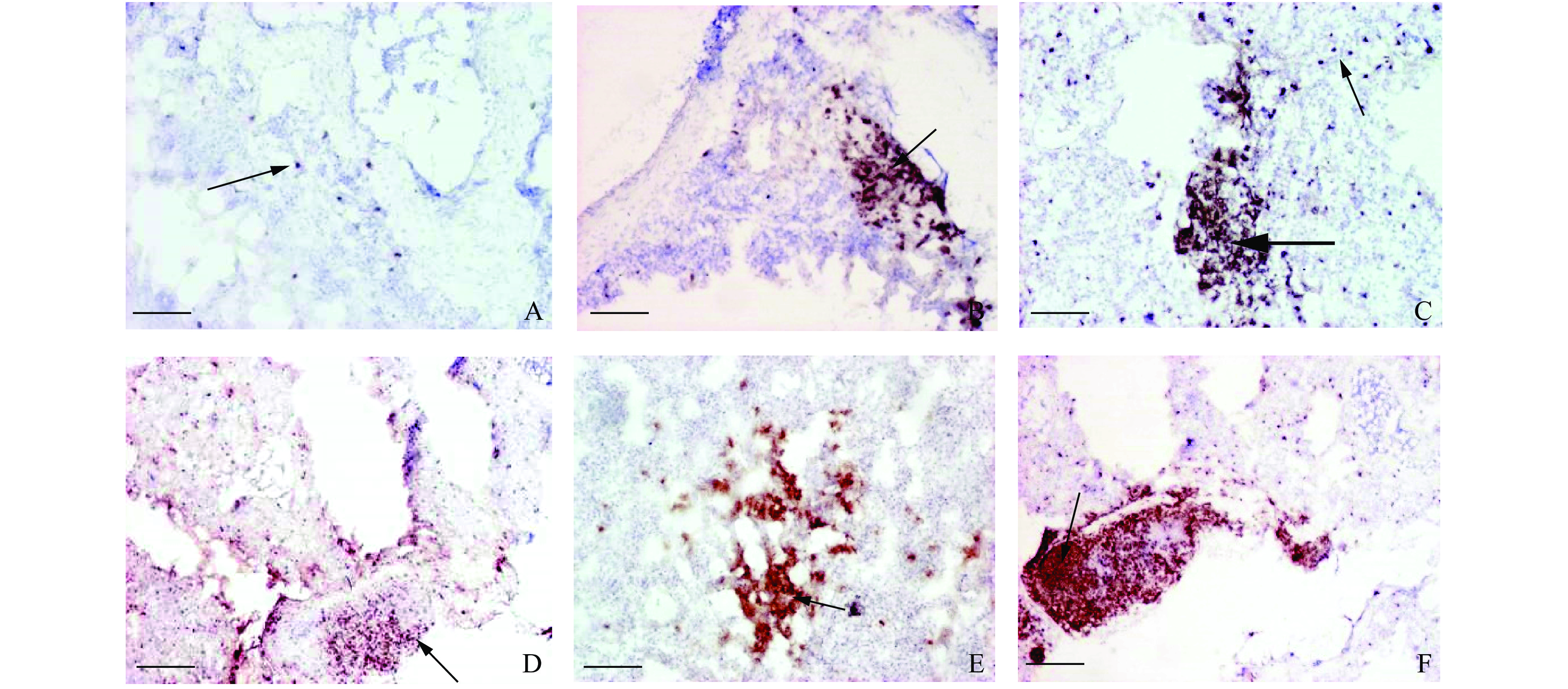
图 3 不同日龄鸡气管黏膜中IgA+分泌细胞的分布情况
A:7日龄(箭头指向气管黏膜固有层中的IgA+细胞),B:14日龄(箭头指向气管黏膜中的IgA+细胞),C:21日龄(箭头指向气管黏膜中的IgA+细胞),D:35日龄(箭头指向黏膜上皮中的IgA+细胞),E:35日龄(箭头指向散布在整个黏膜固有层中的IgA+细胞),F:56日龄(箭头指向生发中心);标尺 = 100 μm
Figure 3. Distribution of IgA+ secreting cells in the tracheal mucosa of chicken at different day-age
A: 7-day-old (arrow points to IgA+ cells in the lamina propria of tracheal mucosa), B: 14-day-old (arrow points to IgA+ cells in the tracheal mucosa), C: 21-day-old (arrow points to IgA+ cells in the tracheal mucosa), D: 35-day-old (arrow points to IgA+ cells in the mucosal epithelium), E: 35-day-old (arrow points to IgA+ cells scattered throughout the lamina propria of mucosa), F: 56-day-old (arrow points to the germinal center); Bar = 100 μm
![]()
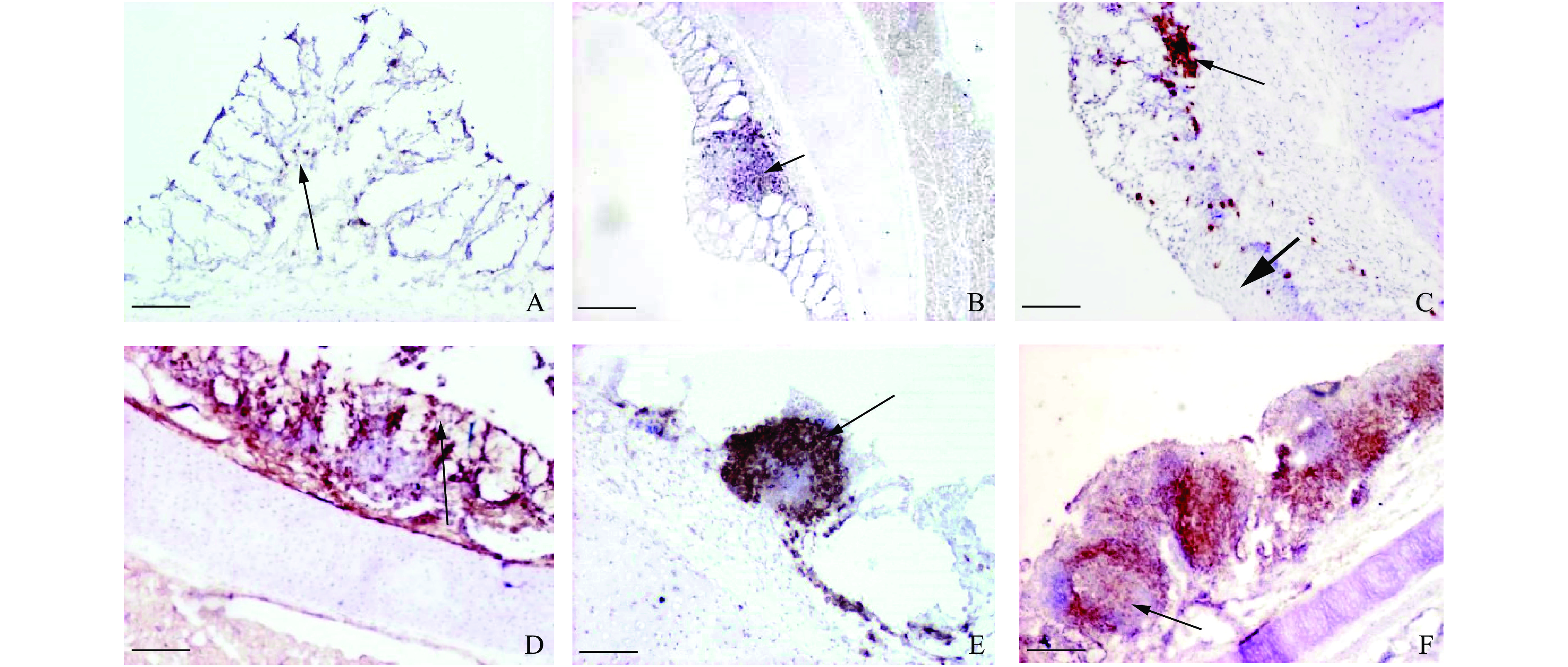
图 4 不同日龄鸡肺脏中IgA+分泌细胞的分布情况
A:1日龄(箭头指向大血管周围的IgA+细胞),B:4日龄(箭头指向支气管相关性淋巴组织中的IgA+细胞),C:14日龄(细箭头指向肺脏实质中的IgA+细胞,粗箭头指向支气管相关性淋巴组织中的IgA+细胞),D:21日龄(箭头指向集中在支气管相关性淋巴组织靠近管腔一侧的IgA+细胞),E:35日龄(箭头指向房间隔上的IgA+细胞),F:56日龄(箭头指向生发中心);标尺 = 100 μm
Figure 4. Distribution of IgA+ secreting cells in the lung of chicken at different day-age
A: 1-day-old (arrow points to IgA+ cells around the great vessels), B: 4-day-old (arrow points to IgA+ cells in the bronchial associated lymphoid tissue), C: 14-day-old (thin arrow points to IgA+ cells in the lung parenchyma; thick arrow points to IgA+ cells in the bronchial associated lymphoid tissue), D: 21-day-old (arrow points to IgA+ cells concentrated near the luminal side of bronchial associated lymphoid tissue), E: 35-day-old (arrow points to IgA+ cells in the atrial septum), F: 56-day-old (arrow points to the germinal center); Bar = 100 μm
-
[1] SMIALEK M, TYKALOWSKI B, STENZEL T, et al. Local immunity of the respiratory mucosal system in chickens and turkeys[J]. Polish Journal of Veterinary Sciences, 2011, 14(2): 291-297.
[2] INVERNIZZI R, LLOYD C M, MOLYNEAUX P L. Respiratory microbiome and epithelial interactions shape immunity in the lungs[J]. Immunology, 2020, 160(2): 171-182. doi: 10.1111/imm.13195
[3] DE GEUS E D, REBEL J M J, VERVELDE L. Induction of respiratory immune responses in the chicken; Implications for development of mucosal avian influenza virus vaccines[J]. Veterinary Quarterly, 2012, 32(2): 75-86. doi: 10.1080/01652176.2012.711956
[4] 张英楠, 杨树宝. 不同日龄蛋鸡肺脏组织结构及其淋巴组织发育的研究[J]. 中国家禽, 2019, 41(6): 11-15. [5] 杨树宝, 冯小刚, 于丹, 等. 鸡鼻黏膜及其淋巴组织发育的组织学观察[J]. 中国兽医杂志, 2014, 50(11): 7-9. doi: 10.3969/j.issn.0529-6005.2014.11.002 [6] 张英楠, 徐晶, 栾维民, 等. 不同日龄鸡肺脏中CD4+、 CD8+ T淋巴细胞分布规律[J]. 中国畜牧兽医, 2019, 46(10): 3052-3057. [7] 张英楠, 杨树宝, 单春兰, 等. 不同日龄蛋鸡气管黏膜中免疫细胞分布的变化规律[J]. 中国兽医科学, 2016, 46(9): 1147-1152. [8] 杨树宝, 张英楠, 马馨, 等. 不同日龄鸡鼻相关性淋巴组织中T和B淋巴细胞分布规律[J]. 中国预防兽医学报, 2014, 36(6): 483-486. doi: 10.3969/j.issn.1008-0589.2014.06.16 [9] 李乙江, 杨晶晶, 牛自兵, 等. 黄牛呼吸道IgA分泌细胞和淋巴组织分布的研究[J]. 南京农业大学学报, 2017, 40(6): 1100-1104. doi: 10.7685/jnau.201609035 [10] FARSTAD I N, CARLSEN H, MORTON H C, et al. Immunoglobulin A cell distribution in the human small intestine: Phenotypic and functional characteristics[J]. Immunology, 2000, 101(3): 354-363. doi: 10.1046/j.1365-2567.2000.00118.x
[11] POTOCKOVA H, SINKOROVA J, KAROVA K, et al. The distribution of lymphoid cells in the small intestine of germ-free and conventional piglets[J]. Developmental and Comparative Immunology, 2015, 51(1): 99-107. doi: 10.1016/j.dci.2015.02.014
[12] 李鹏成, 刘志学, 高君恺, 等. 猪呼吸道IgA和IgG分泌细胞的分布[J]. 畜牧兽医学报, 2010, 41(7): 873-877. [13] VAN GINKEL F W, GULLEY S L, LAMMERS A, et al. Conjunctiva-associated lymphoid tissue in avian mucosal immunity[J]. Developmental and Comparative Immunology, 2012, 36(2): 289-297. doi: 10.1016/j.dci.2011.04.012
[14] KRACKE A, HILLER A S, TSCHERNIG T, et al. Larynx-associated lymphoid tissue (LALT) in young children[J]. Anatomical Record, 1997, 248(3): 413-420. doi: 10.1002/(SICI)1097-0185(199707)248:3<413::AID-AR14>3.0.CO;2-S
[15] LAWRENCE E C, ARNAUD-BATTANDIER F, KOSKI I R, et al. Tissue distribution of immunoglobulin-secreting cells in normal and IgA deficient chickens[J]. Journal of Immunology, 1979, 123(4): 1767-1771.
 下载:
下载:
计量
- 文章访问数: 730
- HTML全文浏览量: 27
- PDF下载量: 835
