
Sequence analysis of AK1 gene from Jingyuan chicken and construction of its eukaryotic expression vector
-
摘要:目的
探究宁夏地方品种静原鸡前期转录组测序筛选出的AK1基因编码蛋白的结构与功能,构建其真核表达载体。
方法根据GenBank上已公布的原鸡AK1基因序列,针对其CDS区设计特异性引物,通过克隆测序对AK1基因CDS区SNPs进行快速筛查,构建AK1基因的真核表达载体,并进行编码区生物信息学功能分析。
结果AK1基因编码区全长585 bp,编码194个氨基酸;AK1蛋白无跨膜结构域,存在2个CpG岛,18个磷酸化位点,10个抗原表位,为稳定的水溶性蛋白,空间结构以α–螺旋和无规则卷曲为主。亚细胞定位结果显示,AK1蛋白主要位于细胞质内;GO富集分析发现,AK1基因同样富集在细胞质中,与亚细胞定位结果一致。基因共表达分析发现,在与AK1互作的基因中,AK1与AMPD1、PKM2基因存在共表达,共表达系数分别为0.116和0.063。成功构建出AK1-pEGFP-N1载体。
结论该研究结果为后续AK1蛋白功能以及AK1作为肌苷酸相关基因的深入研究提供了科学依据。
Abstract:ObjectiveTo explore the structure and function of protein encoded by AK1 gene screened by transcriptome sequencing of Ningxia local breed Jingyuan chicken, and construct its eukaryotic expression vector.
MethodAccording to the original chicken AK1 gene sequence published on GenBank, specific primers were designed for its CDS region, and the CDS region SNPs of AK1 gene were screened quickly by cloning and sequencing. The eukaryotic expression vector of AK1 gene was constructed. The bioinformatics function of the coding region was analyzed.
ResultThe full length of AK1 gene coding region was 585 bp, which encoded 194 amino acids. AK1 protein had no transmembrane domain, and it had two CpG islands, 18 phosphorylation sites and 10 antigenic epitopes. AK1 protein was a stable water-soluble protein, and the spatial structure was mainly α-helix and irregular crimp. The results of subcellular localization showed that AK1 protein was mainly located in the cytoplasm, and GO enrichment analysis showed that AK1 gene was also enriched in the cytoplasm, which was consistent with the result of subcellular localization. Gene co-expression analysis of genes interacting with AK1 showed that AMPD1 and PKM2 coexpressed with AK1 gene, and the co-expression coefficients were 0.116 and 0.063, respectively. The AK1-pEGFP-N1 vector was successfully constructed.
ConclusionThe results of this study provide a scientific basis for further study of the function of AK1 protein and AK1 as an inosinic acid-related gene.
-
Keywords:
- AK1gene /
- bioinformatics /
- Jingyuan chicken /
- gene co-expression /
- subcellular localization
-
-
![]()
图 1 静原鸡不同组织总RNA提取
1、2:胸肌;3、4:腿肌;M:Trans2K Plus DNA Marker
Figure 1. Total RNA extraction from different tissues of Jingyuan chicken
1, 2: Chest muscle; 3, 4: Leg muscle; M: Trans2K Plus DNA Marker
![]()
图 2 静原鸡AK1基因PCR扩增电泳图
M: DL2000 DNA Marker;1~5:扩增片段
Figure 2. Electrophoresis of PCR amplification of AK1 gene in Jingyuan chicken
M: DL2000 DNA Marker; 1–5: Amplified fragments
![]()
图 3 静原鸡AK1基因加酶切位点PCR扩增电泳图
M:DL2000 DNA Marker;1~5:扩增片段
Figure 3. Electrophoresis of PCR amplification of AK1 gene with restriction site in Jingyuan chicken
M: DL2000 DNA Marker; 1–5: Amplified fragments
![]()
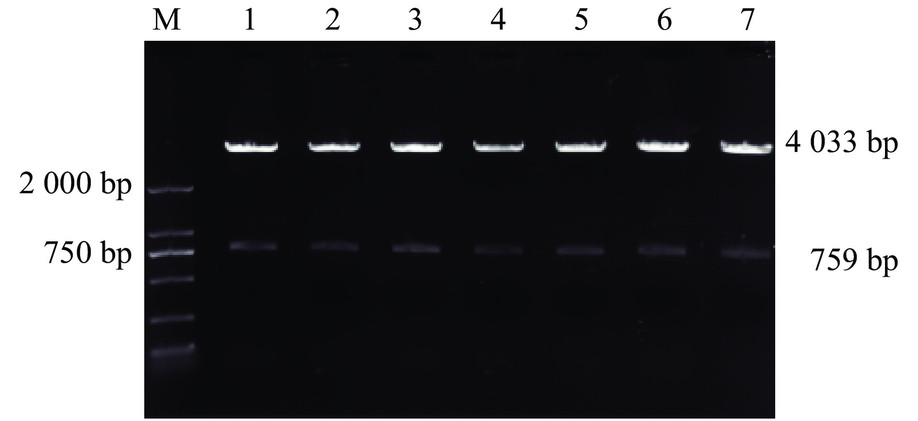
图 4 静原鸡AK1-pEGFP-N1载体酶切电泳图
M:DL2000 DNA Marker;1~7:扩增片段
Figure 4. Restrictive enzyme digestion and electrophoresis of AK1-pEGFP-N1 vector in Jingyuan chicken
M: DL2000 DNA Marker; 1–7: Amplified fragments
![]()
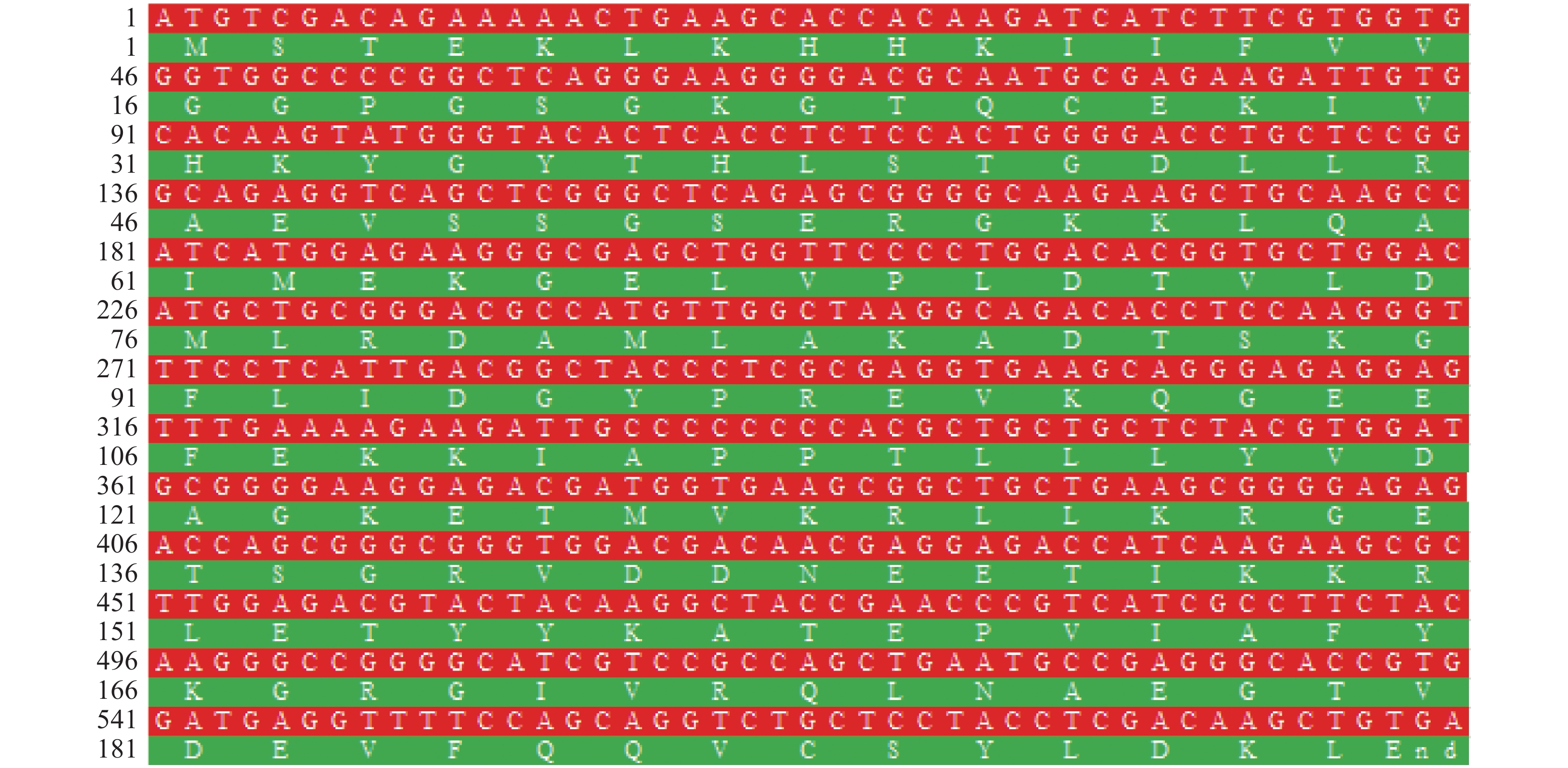
图 5 AK1基因编码区全长序列与对应的氨基酸序列
Figure 5. The full length coding sequence of AK1 gene and corresponding amino acid sequence
![]()
图 7 静原鸡AK1蛋白的疏水性/亲水性分析
Figure 7. Hydrophobic/hydrophilic analysis for AK1 protein in Jingyuan chicken
![]()
图 8 静原鸡AK1蛋白磷酸化位点
Figure 8. The phosphorylation sites of AK1 protein in Jingyuan chicken
![]()
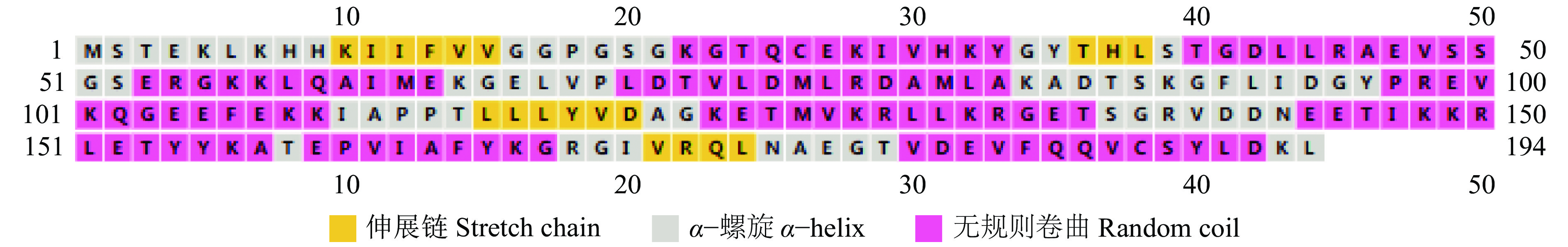
图 10 静原鸡AK1蛋白质二级结构
Figure 10. The secondary structure of AK1 protein in Jingyuan chicken
![]()
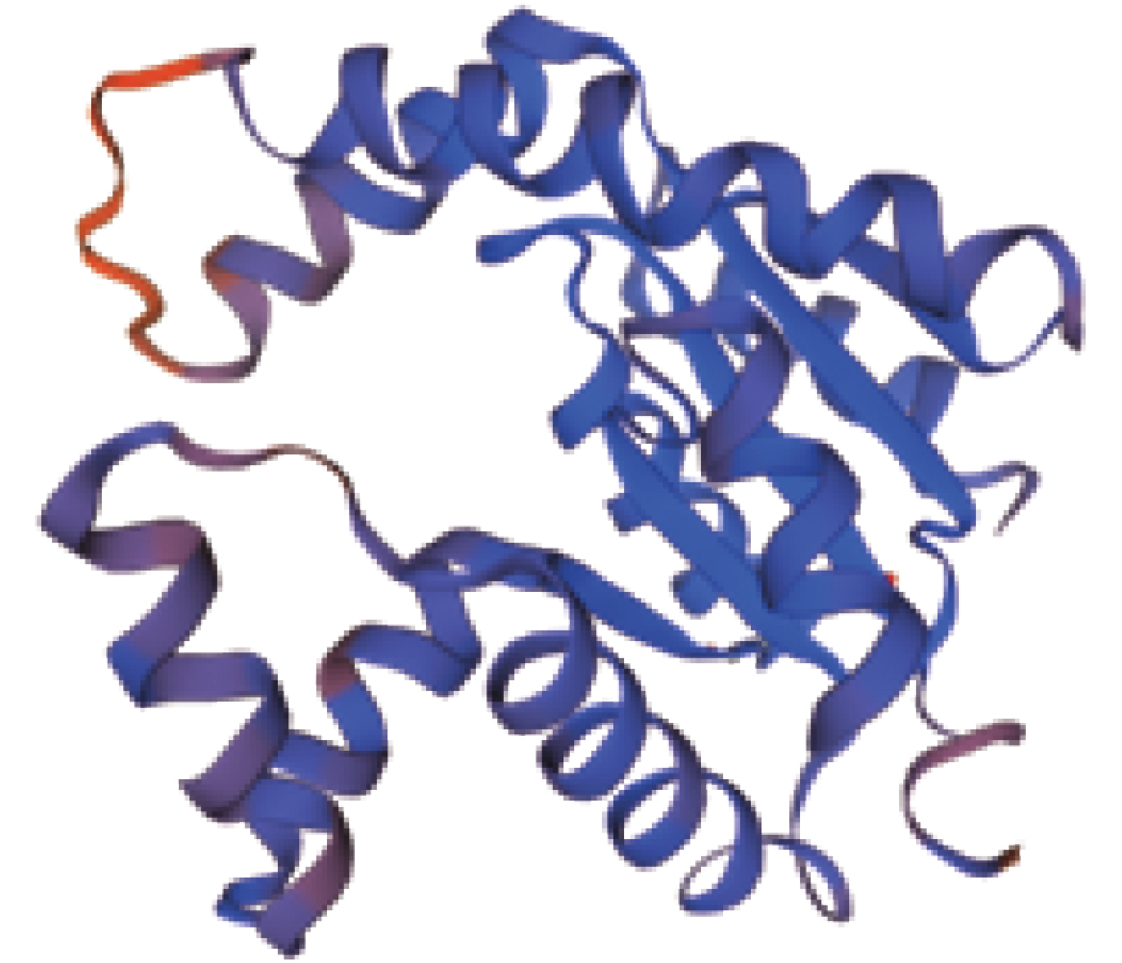
图 11 静原鸡AK1蛋白质预测三级结构
Figure 11. The predicted tertiary structure of AK1 protein in Jingyuan chicken
表 1 静原鸡AK1基因CDS区引物序列
Table 1 Primer sequence for CDS region in AK1 gene of Jingyuan chicken
基因
Gene登录号
Accession number引物序列(5′→3′)1)
Primer sequence产物长度/bp
Product length退火温度/℃
Annealing temperatureAK1 M37901 F:TCCTCCACCCAGACAGCA
R:ACGGGAAAGAGCCAAACA741 57.6 AK13 M37901 F:ccg GAATTCTCCTCCACCCAGACAGCA
R:cgc GGATCCACGGGAAAGAGCCAAACA759 62.7 1) 带有下划线的序列为酶切位点,AK13上、下游引物分别插入EcoRⅠ和BamHⅠ酶切位点
1) Underlined sequences are restriction sites, and forward and reverse primers of AK13 contained EcoR Ⅰ and BamHⅠ restriction sites respectively 下载: 导出CSV
表 2 AK1基因编码蛋白的氨基酸组成
Table 2 The composition of amino acid coded by AK1 gene
氨基酸
Amino Acid数量
Number频率/%
FrequencyAla 10 5.2 Arg 10 5.2 Asn 2 1.0 Asp 11 5.7 Cys 2 1.0 Gln 6 3.1 Glu 18 9.3 Pro 6 3.1 Thr 13 6.7 Val 15 7.7 Gly 19 9.8 His 4 2.1 Ile 9 4.6 Leu 20 10.3 Lys 22 11.3 Met 5 2.6 Phe 5 2.6 Ser 9 4.6 Tyr 8 4.1
下载: 导出CSV
表 3 AK1基因的GO注释分析
Table 3 GO annotation analysis of AK1 gene
项目
ItemGO条目
GO term描述
Description基因数1)
Gene number错误发现率
False discovery rate生物学过程
Biological processGO:0009167 嘌呤核糖核苷单磷酸代谢过程
Purine ribonucleoside monophosphate metabolic process4(41) 2.78×10⁻⁶ GO:0009165 核苷酸生物合成的过程
Nucleotide biosynthetic process4(40) 2.78×10⁻⁶ GO:0009150 嘌呤核糖核苷酸的代谢过程
Purine ribonucleotide metabolic process4(49) 2.78×10⁻⁶ GO:0009168 嘌呤核糖核苷单磷酸生物合成过程
Purine ribonucleoside monophosphate biosynthetic process3(25) 7.56×10⁻⁶ GO:0009152 嘌呤核糖核苷酸的生物合成过程
Purine ribonucleotide biosynthetic process3(29) 8.98×10⁻⁶ GO:0017144 药物代谢过程
Drug metabolic process3(68) 7.47×10⁻⁵ GO:0006188 肌苷酸生物合成过程
IMP biosynthetic process2(7) 8.04×10⁻⁵ GO:0046040 肌苷酸代谢过程
IMP metabolic process2(9) 1.20×10⁻⁴ GO:0006165 二磷酸核苷磷酸化
Nucleoside diphosphate phosphorylation2(14) 2.40×10⁻⁴ GO:0009142 核苷三磷酸生物合成过程
Nucleoside triphosphate biosynthetic process2(21) 4.20×10⁻⁴ GO:0046034 ATP代谢过程
ATP metabolic process2(31) 8.40×10⁻⁴ 分子功能
Molecular functionGO:0003824 催化活性
Catalytic activity5(642) 2.00×10⁻³ GO:0016301 激酶活性
Kinase activity2(98) 7.50×10⁻³ GO:0005524 ATP结合
ATP binding3(202) 7.50×10⁻³ GO:0016787 水解酶活性
Hydrolase activity2(271) 3.13×10⁻² 细胞组分
Cellular componentGO:0005829 细胞溶质
Cytosol3(264) 1.43×10⁻² 1) 括号内的数值表示富集到这个条目的所有基因数,括号外的数值表示差异显著的基因数
1) The value in brackets represents the number of all genes enriched to this GO term, and the value outside brakets represents the number of significantly different genes
下载: 导出CSV
-
[1] 王建保. 朝那鸡列入区级畜禽遗传保护品种[J]. 固原日报, 2009(1): 9-25. [2] 张娟, 母童, 赵平, 等. 静原鸡ELOVL5基因遗传多样性研究[J]. 浙江农业学报, 2019, 31(2): 200-206. doi: 10.3969/j.issn.1004-1524.2019.02.04 [3] 母童, 张娟, 赵平, 等. 静原鸡ELOVL2和ELOVL5基因表达的组织特异性研究[J]. 浙江农业学报, 2017, 29(8): 1290-1296. doi: 10.3969/j.issn.1004-1524.2017.08.09 [4] JONES M E, LIPMANN F. Aceto-CoA-kinase: ATP + CoA + Ac ⇄Ac~S CoA + 5-AMP + PP[J]. Methods in Enzymology, 1955, 1: 585-591. doi: 10.1016/0076-6879(55)01101-4
[5] ZELEZNIKAR R J, HEYMAN R A, GRAEFF R M, et al. Evidence for compartmentalized adenylate kinase catalysis serving a high energy phosphoryl transfer function in rat skeletal muscle[J]. Journal of Biological Chemistry, 1990, 265: 300-311.
[6] VAN ROMPAY A R, JOHANSSON M, KARLSSON A. Phosphorylation of nucleosides and nucleoside analogs by mammalian nucleoside monophosphate kinases[J]. Pharmacolocy & Therapeutics, 2000, 87(2/3): 189-198.
[7] TANABE T, YAMADA M, NOMA T, et al. Tissue- specific and developmentally regulated expression of the genes encoding adenylate kinase isozymes[J]. International Journal of Biochemistry & Cell Biology, 1993, 113(2): 200-207.
[8] SCHULZ G E, ELZINGA M, MARX F, et al. Three-dimensional structure of adenylkinase[J]. Nature, 1974, 250(5462): 120-123. doi: 10.1038/250120a0
[9] SACHSENHEIMER W, SCHULZ G E. Two conformations of crystalline adenylatekinase[J]. Journal of Molecular Biology, 1977, 114(1): 23-36. doi: 10.1016/0022-2836(77)90280-7
[10] HANCOCK C R, JANSSEN E, TERJUNG R L. Skeletal muscle contractile performance and ADP accumulation in adenylate kinase-deficient mice[J]. American Journal of Physiology-Cell Physiology, 2005, 288(6): C1287-C1297. doi: 10.1152/ajpcell.00567.2004
[11] CARRASCO A J, DZEJA P P, ALEKSEEV A E, et al. Adenylate kinase phosphotransfer communicates cellular energetic signals to ATP-sensitive potassium channels[J]. Proceedings of the National Academy of Sciences of the United States of America, 2001, 98(13): 7623-7628. doi: 10.1073/pnas.121038198
[12] 易继财. 生物类专业生物信息学课程教学探索: 华南农业大学生物类专业生物信息学课程的教改实践与思考[J]. 安徽农业科学, 2018, 46(26): 231-233. doi: 10.3969/j.issn.0517-6611.2018.26.071 [13] 吴风瑞, 陈德宇, 姜双林, 等. PBL教学法在硕士研究生生物信息学教学中的应用[J]. 阜阳师范学院学报(自然科学版), 2017, 34(2): 109-111. [14] PANAYIOTOU C, SOLAROLI N, KARLSSON A. The many isoforms of human adenylate kinases[J]. International Journal of Biochemistry & Cell Biology, 2014, 49: 75-83.
[15] JACOBASCH G. Biochemical and genetic basis of red cell enzyme deficiencies[J]. Best Practice & Research Clinical Haematology, 2000, 13(1): 1-20.
[16] ABRUSCI P, CHIARELLI L R, GALIZZI A, et al. Erythrocyte adenylate kinase deficiency: Characterization of recombinant mutant forms and relationship with nonspherocytic hemolytic anemia[J]. Experimental Hematology, 2007, 35(8): 1182-1189. doi: 10.1016/j.exphem.2007.05.004
[17] PUCAR D, BAST P, GUMINA R J, et al. Adenylate kinase AK1 knockout heart: Energetics and functional performance under ischemia-reperfusion[J]. American Journal of Physiology Heart and Circulatory Physiology, 2002, 283(2): H776-H782. doi: 10.1152/ajpheart.00116.2002
[18] SUMINAMI Y, KISHI F, TORIGOE T, et al. Structure and complete nucleotide sequence of the gene encoding chicken cytosolic adenylate kinase[J]. Journal of Biochemistry, 1988, 103(4): 611-617. doi: 10.1093/oxfordjournals.jbchem.a122315
[19] PAI E F, SACHSENHEIMER W, SCHIRMER R H, et al. Substrate positions and induced-fit in crystalline adenylate kinase[J]. Journal of Molecular Biology, 1977, 114(1): 37-45. doi: 10.1016/0022-2836(77)90281-9
[20] FRY D C, KUBY S A, MILDVAN A S. ATP-binding site of adenylate kinase: Mechanistic implications of its homology with ras-encoded p21, F1-ATPase, and other nucleotide-binding proteins[J]. Proceedings of the National Academy of Sciences of the United States of America, 1986, 83(4): 907-911. doi: 10.1073/pnas.83.4.907
[21] GURUPRASAD K, REDDY B V, PANDIT M W. Correlation between stability of a protein and its dipeptide composition: A novel approach for predicting in vivo stability of a protein from its primary sequence[J]. Protein Engineering, 1990, 4(2): 155-161. doi: 10.1093/protein/4.2.155
[22] ZOR T, MAYR B M, DYSON H J, et al. Roles of phosphorylation and helix propensity in the binding of the KIX domain of CREB-binding protein by constitutive (c-myb) and inducible (CREB) activators[J]. Journal of Biological Chemistry, 2002, 277(44): 42241-42248. doi: 10.1074/jbc.M207361200
[23] KYTE J, DOOLITTLE R F. A simple method for displaying the hydropathic character of a protein[J]. Journal of Molecular Biology, 1982, 157(1): 105-132. doi: 10.1016/0022-2836(82)90515-0
[24] 郑海军, 朱荣, 葛春蕾, 等. 人白细胞介素−29的生物信息学分析[J]. 中国生物制品学杂志, 2013, 26(2): 209-212. [25] 赵骁, 张峰波, 王红英, 等. 生物信息分析细粒棘球绦虫EgA31蛋白T细胞及B细胞的优势抗原表位[J]. 中国组织工程研究, 2019, 23(7): 1078-1083. doi: 10.3969/j.issn.2095-4344.1001 [26] CHOO H J, KIM B W, KWON O B, et al. Secretion of adenylate kinase 1 is required for extracellular ATP synthesis in C2C12 myotubes[J]. Experimental and Molecular Medicine, 2008, 40(2): 220-228. doi: 10.3858/emm.2008.40.2.220


计量
- 文章访问数: 942
- HTML全文浏览量: 12
- PDF下载量: 1116
