Effects of azadirachtin on proliferation and apoptosis of Plutella xyllostella embryonic cells
-
摘要:目的
探讨印楝素对小菜蛾Plutella xyllostella胚胎细胞增殖和凋亡的影响及可能的机制。
方法将体外培养的小菜蛾胚胎细胞分为印楝素处理组和未处理组,采用CCK-8试剂盒检测细胞增殖的抑制率,激光共聚焦镜检PI染色后的细胞死亡和DAPI染色后的凋亡小体,通过Western-blotting检测各组细胞Caspase-3的表达情况以及通路蛋白Akt的磷酸化水平。
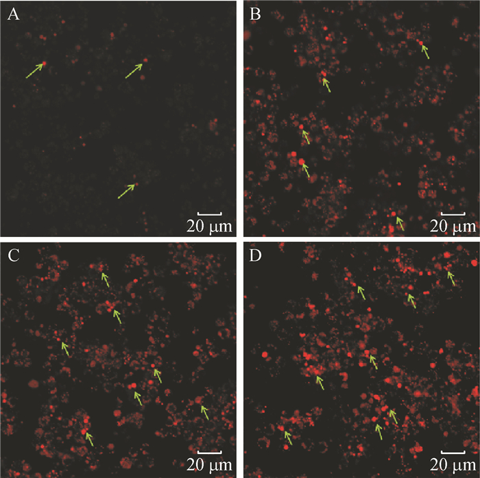
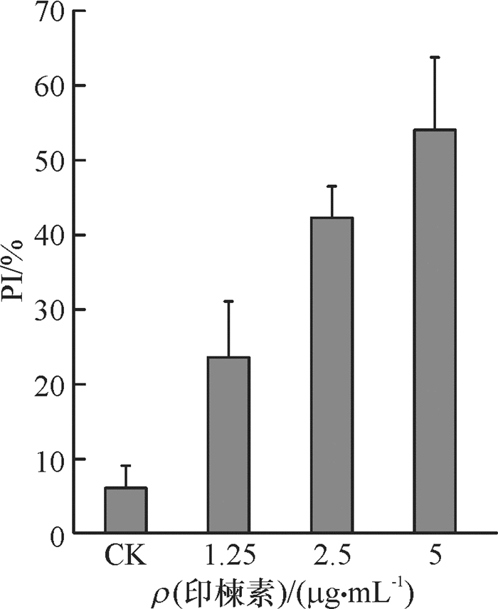
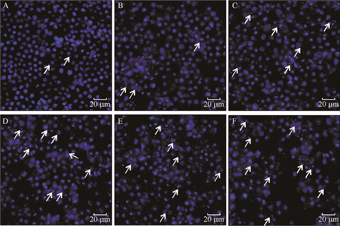
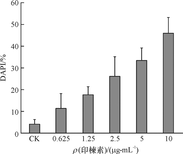
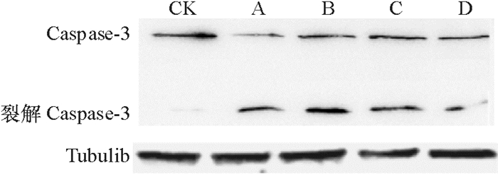
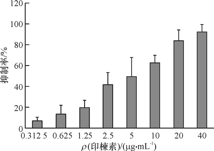
结果印楝素对小菜蛾胚胎细胞有明显的增殖抑制作用,且呈现浓度依赖性,24 h的IC50为4.4 μg·mL-1。印楝素处理后的细胞经PI染色后能明显观察到死亡细胞,DAPI染色后可见凋亡小体;Caspase-3蛋白发生剪切,并且抑制Akt的磷酸化水平。
结论印楝素对小菜蛾胚胎细胞的增殖有明显的抑制作用,并通过抑制Akt信号通路的活化,诱导细胞产生依赖于Caspase-3的Ⅰ型凋亡。
Abstract:ObjectiveTo investigate the effect and mechanism of azadirachtin on proliferation and apoptosis in Plutella xyllostella embryonic cells.
MethodP. xyllostella embryonic cells were divided into azadirachtin treated and untreated groups, the inhibition rate of cells proliferation was detected using CCK-8 kit. The cell death and apoptotic body were observed using laser confocal microscope after stained by PI and DAPI respectively. The expression of Caspase-3 and the phosphorylation of pathway protein Akt were detected using Western-blotting.
ResultAzadirachtin had significant inhibitory effect on the proliferation of P. xyllostella embryonic cells, and the effect was dose-dependent. IC50 was 4.4 μg·mL-1 in 24 h. Dead cells could be observed in azadirachtin treated group after stained by PI, and apoptotic body could be found in the same group after stained by DAPI. Caspase-3 protein was cleaved and Akt phosphorylation was inhibited.
ConclusionAzadirachtin has significant inhibitory effect on the proliferation of P. xyllostella embryonic cells. By inhibiting the activation of Akt signaling pathway, azadirachtin can induce cells to produce Caspase-3-dependent type I apoptosis.
-
Keywords:
- azadirachtin /
- Plutella xyllostella /
- embryonic cell /
- apoptosis /
- proliferation
-
-
-
[1] 张晓晖, 姚天明, 黄高昇, 等.细胞凋亡的最新研究进展[J].第四军医大学学报, 2002, 23(12): 42-44. http://youxian.cnki.com.cn/yxdetail.aspx?filename=YAOL20160620000&dbname=CAPJ2015 [2] NEZHA F, COHEN C, RAHAMAN J, et al. Comparative immunohistochemical studies of bcl-2 and P53 proteins in benign and malignant ovarian endometriotic cysts[J]. Cancer, 2002, 94(11): 2935-2940. doi: 10.1002/(ISSN)1097-0142
[3] NASU K, NISHIDA M, KAWANO Y, et al. Aberrant expression of apoptosis-related molecules in endometriosis: A possible mechanism underlying the pathogenesis of endometriosis[J]. Rerrod Sci, 2011, 18(3): 206-218. doi: 10.1177/1933719110392059
[4] IMMARAJU J A. The commercial use of azadirachtin and its integration into viable pest control programmes[J]. Pestic Sci, 2015, 54(3): 285-289. http://cat.inist.fr/?aModele=afficheN&cpsidt=2428852
[5] VASSILIOU V A. Botanical insecticides in controlling Kelly's citrus thrips (Thysanoptera: Thripidae) on organic grapefruits[J]. J Econ Entomol, 2011, 104(6): 1979-1985. doi: 10.1603/EC11105
[6] SRIVASTAVA S, SRIVASTAVA A K. In vitro azadirachtin production by hairy root cultivation of azadirachta indica in nutrient mist bioreactor[J]. Appl Biochem Biotech, 2012, 166(2): 365-378. doi: 10.1007/s12010-011-9430-9
[7] 李文欧, 徐汉虹, 张志祥, 等.印楝素对粉纹夜蛾Hi-5细胞的毒性机理[J].昆虫学报, 2008, 51(8): 824-829. http://www.cnki.com.cn/Article/CJFDTOTAL-KCXB200808006.htm [8] 钟国华, 水克娟, 吕朝军, 等.印楝素对SL1的细胞凋亡诱导作用[J].昆虫学报, 2008, 51(6): 618-627. http://www.cnki.com.cn/Article/CJFDTOTAL-KCXB200806010.htm [9] 程杏安, 黄劲飞, 胡美英, 等.印楝素诱导Sf9细胞凋亡的显微和超微形态变化[J].华南农业大学学报, 2010, 31(4): 52-58. http://xuebao.scau.edu.cn/zr/hnny_zr/ch/reader/view_abstract.aspx?file_no=201004104&flag=1 [10] HUANG J F, SHUI K J, LI H Y, et al. Antiproliferative effect of azadirachtin on Spodoptera litura Sl-1 cell line through cell cycle arrest and apoptosis induced by up-regulation of p53[J]. Pestic Biochem Phys, 2011, 99(1): 16-24. doi: 10.1016/j.pestbp.2010.08.002
[11] WANG Z, CHENG X G, MENG Q Q, et al. Azadirachtin-induced apoptosis involves lysosomal membrane permeabilization and cathepsin L release in Spodoptera frugiperda Sf9 cells[J]. Int J Biochem Cell B, 2015, 64: 126-135. doi: 10.1016/j.biocel.2015.03.018
[12] SHAO X H, LAI D, ZHANG L, et al. Induction of autophagy and apoptosis via PI3K/AKT/TOR pathways by azadirachtin A in Spodoptera litura cells[J]. Sci Rep-UK, 2016, 6: 35482. doi: 10.1038/srep35482
[13] FISHEL F M. IRAC's insecticide mode of action classification[M]. Florida: Florida Cooperative Extension Service, Institute of Food and Agricultural Sciences, University of Florida, 2008: 1-5.
[14] WANG H, LAI D, YUAN M, et al. Growth inhibition and differences in protein profiles in azadirachtin-treated Drosophila larvae[J]. Electrophoresis, 2014, 35(8): 1122-1129. doi: 10.1002/elps.v35.8
[15] WEI W, GAI Z, AI H, et al. Baculovirus infection triggers a shift from amino acid starvation-induced autophagy to apoptosis[J]. PLoS One, 2012, 7(5): e37457. doi: 10.1371/journal.pone.0037457
[16] WU F, LI Y, WANG F, et al. Differential function of the two Atg4 homologues in the aggrephagy pathway in Caenorhabditis elegans[J]. J Biol Chem, 2012, 287(35): 29457-29467. doi: 10.1074/jbc.M112.365676
[17] 李小明, 孙志贤.细胞凋亡中的关键蛋白酶Caspase-3[J].医学分子生物学杂志, 1999, 1: 6-9. http://youxian.cnki.com.cn/yxdetail.aspx?filename=ZXDY20171016005&dbname=CAPJ2015 [18] ROSEN N, SHE Q B. Akt and cancer is it all mTOR?[J]. Cancer Cell, 2006, 10(4): 254-256. doi: 10.1016/j.ccr.2006.10.001
[19] XIN M G, DENG X M. Nicotine inactivation of the proapoptotic function of bax through phosphorylation[J]. J Biol Chem, 2005, 280(11): 10781-10789. doi: 10.1074/jbc.M500084200
[20] SONG G, OUYANG G L, BAO S D. The activation of Akt/PKB signaling pathway and cell survival[J]. J Cell Mol Med, 2005, 9(1): 59-71. doi: 10.1111/jcmm.2005.9.issue-1
[21] GIBSON E M, HENSON K S, HANEY N, et al. Epidermal growth factor protects epithelial-derived cells from tumor necrosis factor-related apoptosis-inducing ligand-induced apoptosis by inhibiting cytochrome c release[J]. Cancer Res, 2002, 62(2): 488-496. https://www.ncbi.nlm.nih.gov/pubmed/%20%20%20%20%20%2011809700
[22] KIM E C, YUN B S, RYOO I J, et al. Complestatin prevents apoptotic cell death: Inhibition of a mitochondrial caspase pathway through AKT/PKB activation[J]. Biochem Bioph Res Co, 2004, 313(1): 193-204. doi: 10.1016/j.bbrc.2003.11.104


 下载:
下载:
