Mechanism and influence of down-regulation of miR-29a-3p expression on apoptosis in mouse macrophage
-
摘要:目的
研究miR-29a-3p下调表达对小鼠巨噬细胞凋亡及相关基因表达的影响,为探讨结核病发病机制、研发新的诊断与治疗方法提供依据。
方法将重组抑制载体pEZX-AM02-miR-29a-3p转染RAW264.7细胞,24、36和48 h荧光显微镜观察转染效率,Real time-PCR法检测miR-29a-3p及凋亡相关基因caspase3、caspase7、caspase8、Bcl-2、Mcl-1和Bax的表达水平,流式细胞术检测RAW264.7细胞的凋亡率。
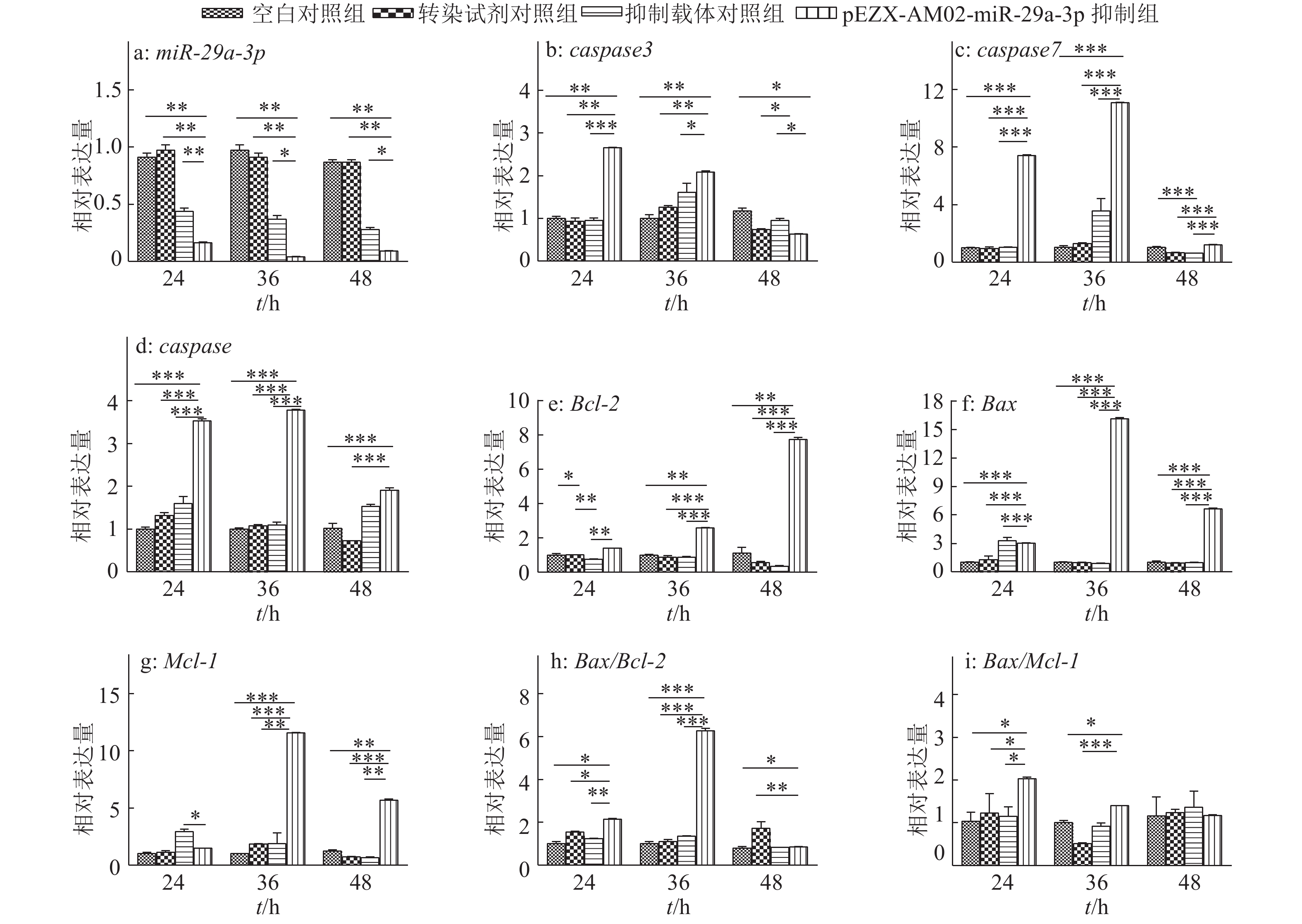
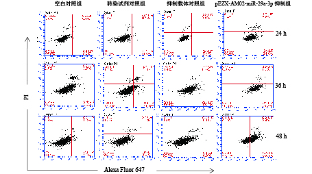
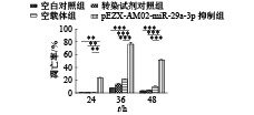
结果重组抑制载体转染后,细胞内miR-29a-3p表达水平明显下降;而caspase7、caspase8、Bcl-2、Mcl-1和Bax等基因的表达出现不同程度的上调;流式细胞术分析发现随着转染时间延长,细胞凋亡率增加,且36 h凋亡率最高。
结论pEZX -AM02-miR-29a-3p重组载体可抑制小鼠巨噬细胞中miR-29a-3p的表达,通过靶向上调caspase7、caspase8、Bcl-2和Mcl-1等基因的表达促进巨噬细胞的凋亡。
-
关键词:
- miR-29a-3p /
- 抑制载体 /
- 巨噬细胞 /
- 细胞凋亡 /
- 靶基因
Abstract:ObjectiveTo study the influence of down-regulation of miR-29a-3p expression on apoptosis in mouse macrophage and expression of apoptosis-associated genes, and provide references for investigating the pathogenesis mechanism of mycobacterium tuberculosis (MTB) and developing new diagnosis and treatment methods.
MethodThe recombinant inhibitor vector pEZX-AM02-miR-29a-3p was transfected into RAW264.7 cells. Transfection efficiency was observed through fluorescence microscopy, the expression levels of miR-29a-3p and apoptosis-associated genes caspase3, caspase7, caspase8, Bcl-2, Mcl-1 and Bax were detected by RT-PCR, and the apoptosis rates of RAW264.7 cells were detected by flow cytometry at 24, 36, 48 h after transfection.
ResultAfter transfected with pEZX-AM02-miR-29a-3p recombinant inhibitor vector, miR-29a-3p expression in RAW264.7 cells decreased significantly, while the target genes caspase7, caspase8, Bcl-2, Mcl-1 and Bax were up-regulated at different levels. The results of flow cytometry indicated that the apoptosis rates increased as the transfection time progressed and reached the peak at 36 h.
ConclusionThe expression of miR-29a-3p in mouse macrophage are inhibited by transfecting with pEZX-AM02-miR-29a-3p, which promotes the apoptosis of macrophage by up-regulating the expression of target genes, suah as caspase7, caspase8, Bcl-2 and Mcl-1.
-
Keywords:
- miR-29a-3p /
- inhibitor vector /
- macrophage /
- cell apoptosis /
- target gene
-
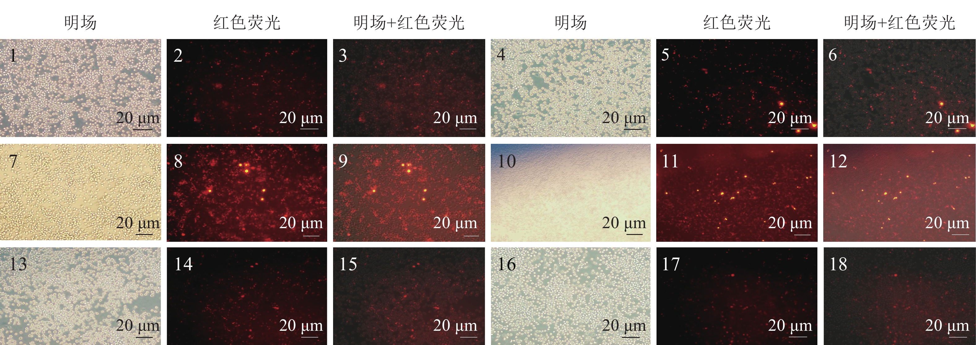
图 1 miRNA抑制对照以及pEZX-AM02-miR-29a-3p转染RAW264.7细胞
1~3为24 h的抑制载体对照(pEZX-AM02);4~6为24 h的pEZX-AM02-miR-29a-3p载体;7~9为36 h的抑制载体对照(pEZX-AM02);10~12为36 h的pEZX-AM02-miR-29a-3p载体;13~15为48 h的抑制载体对照(pEZX-AM02);16~18为48 h的pEZX-AM02-miR-29a-3p载体
Figure 1. RAW264.7 cells tranfected with miRNA inhibitor control or pEZX-AM02-miR-29a-3p
表 1 基因引物序列
Table 1 Sequences of gene primers
基因 上游引物(5'→3') 下游引物(5'→3') caspase3 TCTGACTGGAAAGCCGAAAC GCAAGCCATCTCCTCATCA caspase7 AAACCCTGTTAGAGAAACCCAA TAAGCAAAGAGGAAGTCGGC caspase8 GCTGCCCTCAAGTTCCTGT GATTGCCTTCCTCCAACATC Bcl-2 GGTGGAGGAACTCTTCAGGG ACATCTCCCTGTTGACGCTC Bax ATGCGTCCACCAAGAAGC CAGTTGAAGTTGCCATCAGC Mcl-1 TGTAAGGACGAAACGGGACT CAAAAGCCAGCAGCACATT -
[1] GRIFFITHS-JONES S, GROCOCK R J, VAN DONGEN S, et al. miRBase: microRNA sequences, targets and gene nomenclature[J]. Nucleic Acids Res, 2006, 34: 140-144.
[2] O’CONNELL R M, RAO D S, CHAUDHURI A A, et al. Physiological and pathological roles for microRNAs in the immune system[J]. Nat Rev Immunol, 2010, 10(2): 111-22.
[3] FENTON M J, VERMEULEN M W. Immunopathology of tuberculosis: Roles of macrophages and monocytes[J]. Infect Immun, 1996, 64(3): 683-690.
[4] AUGENSTREICH J, ARBUES A, SIMEONE R, et al. ESX-1 and phthiocerol dimycocerosates of Mycobacterium tuberculosis act in concert to cause phagosomal rupture and host cell apoptosis[J]. Cell Microbiol, 2017, 19(7): e12726.
[5] GUPTA A, KAUL A, TSOLAKI A G, et al. Mycobacterium tuberculosis: Immune evasion, latency and reactivation[J]. Immunobiology, 2012, 217(3): 363-374.
[6] LIU M, WU L, XIANG X, et al. Mycobacterium tuberculosis, effectors interfering host apoptosis signaling[J]. Apoptosis, 2015, 20(7): 883-891.
[7] 包孟, 付玉荣, 伊正君. 微小RNA在机体抗结核免疫及结核诊断中的研究进展[J]. 中华结核和呼吸杂志, 2015, 38(12): 918-921. [8] DAS K, SAIKOLAPPAN S, DHANDAYUTHAPANI S. Differential expression of miRNAs by macrophages infected with virulent and avirulent Mycobacterium tuberculosis[J]. Tuberculosis, 2013, 93(Suppl): 47-50.
[9] 李金妹. 不同分枝杆菌侵染对巨噬细胞免疫相关基因及microRNAs表达的影响[D]. 长春: 吉林农业大学, 2015. [10] SHARBATI J, LEWIN A, KUTZLOHROFF B, et al. Integrated microRNA-mRNA-analysis of human monocyte derived macrophages upon Mycobacterium avium subsp. hominissuis infection[J]. Plos One, 2011, 6(6): e20258.
[11] 董伟杰, 李微, 刘丹霞, 等. 不同毒力结核分枝杆菌感染对巨噬细胞caspase-3及Bcl-2表达的影响[J]. 中国病原生物学杂志, 2013(4): 302-306. [12] SLY L M, HINGLEYWILSON S M, REINERr N E, et al. Survival of Mycobacterium tuberculosis in host macrophages involves resistance to apoptosis dependent upon induction of antiapoptotic Bcl-2 family member Mcl-1[J]. J Immunol, 2003, 170(1): 430-437.
[13] KOZIEL J, MACIAGGUDOWSKAN A, MIKOLAJCZYK T, et al. Phagocytosis of Staphylococcus aureus by macrophages exerts cytoprotective effects manifested by the upregulation of antiapoptotic factors[J]. PLoS One, 2009, 4(4): e5210.
[14] 刘云霞, 张万江. 结核分枝杆菌与巨噬细胞相互作用的研究进展[J]. 中国细胞生物学学报, 2012(6): 617-622. [15] 马烽. microRNA介导的IL-10与IFN-γ表达的转录后调控机制研究[D]. 杭州: 浙江大学, 2011. [16] 杨静, 董剑. miRN A与结核分枝杆菌感染的研究进展[J]. 重庆医学, 2015(8): 1132-1134. [17] RAJARAM M V, NI B, DODD C E, et al. Macrophage immunoregulatory pathways in tuberculosis[J]. Semin Immumol, 2014, 26(6): 471-485.


 下载:
下载:


